Talk to Lawyer
Mark Kopec Now
Mark Kopec Now
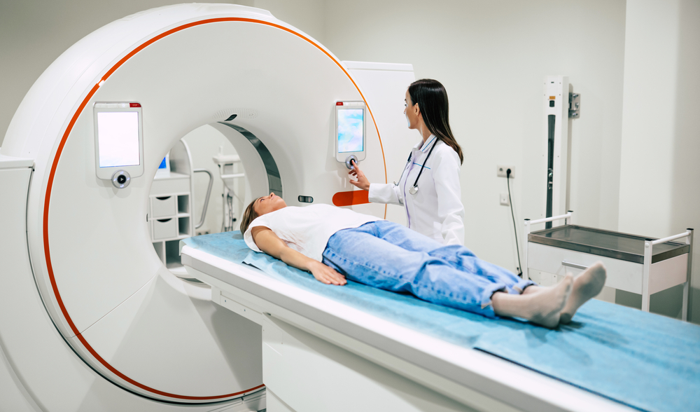
Positron Emission Tomography (PET) scans represent one of the most sophisticated and powerful diagnostic tools in modern medicine. It offers an unparalleled view into the metabolic function of the human body. Unlike traditional imaging that captures anatomy, PET captures activity. As a result, it is invaluable for detecting disease at its earliest, most active stages. However, like any advanced technology, PET scan’s complex nature introduces unique risks for misinterpretation and procedural errors. Pet scans are directly at the heart of medical malpractice litigation.
The conceptual foundation for PET scanning dates back over a century, long before the technology was practical. The notion that blood flow and brain function are intrinsically linked was first appreciated in the late 1800s. However, the true development of PET began with the discoveries of the atomic age.
Key milestones include:
A doctor orders a PET scan when they need to observe the physiological activity or metabolism of tissues and organs, rather than just their structure.
PET scans are primarily in three major medical fields: Oncology, Cardiology, and Neurology.
| Field | Purpose and Circumstances |
| Oncology (Cancer) | The most common use. PET is vital for: |
| Diagnosis and Staging: Determining if a tumor is malignant (identifying “hot spots” of high glucose uptake). | |
| Metastasis Detection: Finding if cancer has spread to lymph nodes or distant organs. | |
| Treatment Monitoring: Assessing if chemotherapy or radiation is effective (successful treatment results in reduced FDG uptake). | |
| Recurrence Surveillance: Detecting if cancer has returned after treatment. | |
| Cardiology (Heart Disease) | To assess heart function and blood flow: |
| Myocardial Viability: Determining which parts of the heart muscle are scarred (dead) versus merely stunned (hibernating) and potentially salvageable through intervention (like bypass surgery). | |
| Neurology (Brain Disorders) | To examine brain metabolism and function: |
| Dementia: Diagnosing and differentiating types of dementia, such as Alzheimer’s disease (which shows characteristic patterns of reduced FDG uptake in specific brain regions). | |
| Epilepsy: Precisely locating the seizure focus in patients who do not respond to medication and are candidates for surgery. | |
| Brain Tumors: Aiding in the grading and biopsy planning of masses. |
PET scans are specialized procedures ordered by doctors who manage complex diseases.
Often, doctors use PET scans in conjunction with, and compared to, Computed Tomography (CT Scan) and Magnetic Resonance Imaging (MRI), as the three modalities offer different, yet complementary, information.
| Modality | Basis of Image | What It Shows (Strengths) | What It Cannot Show (Weaknesses) |
| PET Scan | Function/Metabolism (Radiotracer Uptake) | Cellular activity; early disease; spread of cancer (staging); treatment response. | Poor spatial resolution; limited anatomical detail. |
| CT Scan | Anatomy/Structure (X-rays) | Dense tissue (bone); precise size and location of masses; trauma/hemorrhage; lung tissue. | Poor soft tissue and neurological detail; uses ionizing radiation. |
| MRI Scan | Anatomy/Structure (Magnets and Radio Waves) | Superior soft-tissue contrast (brain, spinal cord, ligaments, joints). | Not ideal for bone/lung; longer scan time; absolute contraindications for metal implants (pacemakers, certain clips). |
| PET/CT (Hybrid) | Function + Anatomy | Combines the strengths: pinpointing the metabolic “hot spot” (PET) onto the exact anatomical location (CT). | Higher radiation dose (from both CT and PET); cost. |
A PET scan’s strength lies in its ability to detect physiological changes before they result in visible anatomical changes on a CT or MRI. Conversely, PET scans can have false positives (high metabolic activity in benign conditions like inflammation or infection) or false negatives (cancers that are slow-growing or use a non-glucose metabolic pathway).
PET scans can be involved in a medical malpractice claim in two distinct ways: by being the source of the alleged negligence or by serving as critical evidence to prove negligence.
Negligence related to PET scans typically falls into one of three categories:
In litigation, the PET scan is often the most important piece of evidence.
In summary, the PET scan, while a powerful life-saving technology, introduces layers of complexity where diagnostic judgment and inter-specialty communication are paramount. Interpreting and acting upon these functional images requires high standards of care. When breached, the result is often a costly and medically damaging diagnostic delay that forms the basis of a medical malpractice lawsuit.
If you have concerns about PET scans and medical malpractice, visit our free consultation page or video. Then contact the Kopec Law Firm at 800-604-0704 to speak directly with Attorney Mark Kopec. He is a top-rated Baltimore medical malpractice lawyer. The Kopec Law Firm is in Baltimore and pursues cases throughout Maryland and Washington, D.C.