Talk to Lawyer
Mark Kopec Now
Mark Kopec Now


The birth of a child is one of the most profound moments in human life. But it is also a complex physiological process fraught with risk. During labor, a fetus must endure the stress of uterine contractions. They temporary restrict oxygen rich blood flow through the placenta. Many babies tolerate this process well. However, some experience fetal distress. This was a critical shortage of oxygen that can lead to permanent injury or death if left unmanaged. An important player in these situations is fetal heart rate monitoring.
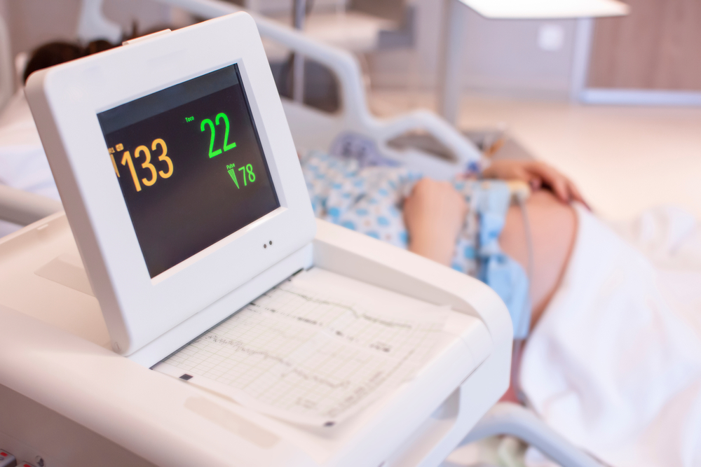
To detect these hidden crises, obstetric medicine relies heavily on Electronic Fetal Monitoring (EFM). As a pillar of modern labor management, fetal heart monitoring provides a continuous window into the baby’s well-being. Interpreting these readings requires a high degree of skill. As a result, it is also one of the most frequent subjects for medical malpractice cases. When medical providers fail to accurately read a fetal heart strip or delay necessary interventions, the consequences can be a birth injury that is catastrophic and lifelong.
Before the mid-20th century, assessing a fetus during labor was intermittent and used low technology. Obstetricians and midwives used a specialized stethoscope called a Pinard horn (invented in the 19th century) or a standard stethoscope to listen to the fetal heartbeat at intervals between contractions.
The fundamental purpose of fetal heart monitoring is to detect fetal hypoxia (oxygen deprivation) and ischemia (inadequate blood flow) before they cause permanent brain damage or death.
During a uterine contraction, it compresses the blood vessels giving the placenta, temporary reducing the delivery of oxygen rich blood to the fetus. A healthy fetus with adequate placental reserves can easily tolerate this temporary dip. However, if the placenta is compromised, the umbilical cord is compressed, or there is prolonged labor, the fetus may deplete its oxygen reserves. Fetal heart monitoring tracks how the baby’s heart reacts to these stresses, allowing providers to tell between a baby who is safely coping and one who is decompensating.
Fetal monitoring occurs in two primary clinical contexts:
Performed in the third trimester (usually after 28 weeks) for high-risk pregnancies, such as those involving maternal hypertension, gestational diabetes, twins, or suspected fetal growth restriction (IUGR). Common tests include:
This is the most common usage. It begins upon admission to the labor and delivery unit and continues until the baby is born. Monitoring can be intermittent (listening at scheduled intervals for low-risk pregnancies) or continuous (mandatory for high-risk pregnancies, induced labors, or when epidural anesthesia is utilized).
Fetal monitoring is a collaborative, multidisciplinary responsibility. The primary medical providers involved include:
Medical providers use two primary categories to monitor a fetus:
This is the most common method and utilizes two separate discs (transducers) secured to the mother’s abdomen with elastic bands:
If the external trace is unclear (often due to maternal movement or tissue thickness) or if labor is failing to progress, providers may switch to internal monitoring. This requires the amniotic sac to be ruptured and the cervix to be partially dilated:
To interpret a fetal heart strip, clinicians look at four core components defined by the National Institute of Child Health and Human Development (NICHD). Medical malpractice cases often hinge on whether providers documented and reacted to these specific metrics:
The medical community categorizes fetal heart rate tracings into a three-tier system to standardize care and minimize subjective errors.
| Category | Clinical Meaning | Interpretation & Characteristics |
|---|---|---|
| Category I | Normal | Strongly predictive of normal fetal acid-base status. Requires routine care. • Baseline: 110–160 bpm • Variability: Moderate • Late/Variable Decelerations: Absent • Early Decelerations: Present or absent |
| Category II | Indeterminate | Not predictive of abnormal acid-base status, but requires continued evaluation, surveillance, and reevaluation. Includes a wide mix of tracings (e.g., minimal variability, marked variability, prolonged decelerations). |
| Category III | Abnormal | Predictive of abnormal fetal acid-base status. Requires prompt evaluation and immediate corrective action. Includes: • Absent variability plus recurrent late decelerations, recurrent variable decelerations, or bradycardia. • Sinusoidal heart rate pattern. |
When a fetal heart strip shifts into Category II or Category III, the standard of care requires providers to take immediate action. The initial steps are referred to as intrauterine resuscitation techniques:
Delivery Intervention: If intrauterine resuscitation fails to resolve a Category III tracing, or if the tracing reveals a prolonged, non-recovering deceleration (bradycardia), the standard of care dictates an immediate operative delivery, typically via an emergency C-section. In emergency obstetrics, the unofficial benchmark is the “30-minute rule”—the time from the decision to operate to the actual incision and delivery of the baby should ideally not exceed 30 minutes.
When medical personnel fail to recognize or properly act on signs of fetal distress, the baby’s brain is progressively starved of oxygen. This can lead to a cascade of catastrophic injuries:
Fetal heart monitoring sits at the center of many birth injury lawsuits. Malpractice claims rarely argue that the doctor or nurse caused the underlying umbilical cord twist or placental failure; rather, they argue that the providers failed to diagnose and treat the distress in a timely manner. Common categories of these claims include:
This occurs when an L&D nurse or physician misinterprets a Category II or III strip as reassuring (Category I). For example, mistaking a dangerous late deceleration for a benign early deceleration, or failing to notice that fetal heart rate variability has shifted from moderate to completely absent.
Labor and delivery units operate under a strict hierarchical chain of command. If an L&D nurse identifies a worsening, dangerous fetal heart rate tracing and notifies the attending obstetrician, but the doctor fails to respond or dismisses the concern, the nurse has a legal duty to bypass that doctor. This means calling the charge nurse, the department head, or the medical director. Failure by the nursing staff to advocate for the patient through the chain of command is a frequent basis for hospital liability.
Even if the team correctly identifies fetal distress, liability arises if they delay the delivery. This can occur due to an unavailable operating room, delays in securing an anesthesia provider, or an obstetrician waiting too long in hopes that the tracing will spontaneously improve. If those minutes of delay cause the baby to cross the threshold into permanent brain injury, medical malpractice may have occurred.
Pitocin is a highly potent medication. If a nurse continues to increase or fails to stop Pitocin when the monitor shows tachysystole (more than 5 contractions in a 10-minute window) or when non-reassuring heart rate patterns emerge, it may constitute breach of the standard of care. The excessive contractions effectively suffocate the fetus, and continuing the medication under those conditions is a frequent cause of successful medical malpractice lawsuits.
Electronic fetal monitoring is a valuable shield against birth trauma, but its value relies entirely on the human eyes reading the printout. When clinical malpractice blinds providers to the clear warnings on a fetal monitor strip, the law provides a pathway for families to seek accountability and secure the substantial financial resources required to care for a permanent injured child.
You can read Baltimore Medical Malpractice Lawyer Blog posts on birth injury verdicts involving fetal heart rate monitoring:
If you have concerns about fetal heart rate monitoring and birth injury medical malpractice, visit our free consultation page or video. Then contact the Kopec Law Firm at 800-604-0704 to speak directly with Attorney Mark Kopec. He is a top-rated Baltimore medical malpractice lawyer. The Kopec Law Firm is in Baltimore and pursues cases throughout Maryland and Washington, D.C.