Talk to Lawyer
Mark Kopec Now
Mark Kopec Now


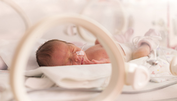
The birth of a child is a complex medical event. It requires precise monitoring, sound clinical judgment, and following of accepted standards of care. While many deliveries proceed without things going wrong, the mechanical forces involved in birth can sometimes cause birth injuries. One visible sign of birth trauma is a subconjunctival hemorrhage. It is the rupture of tiny, fragile blood vessels beneath the clear surface of the baby’s eye. If your child has been injured, you may need Baltimore subconjunctival hemorrhage lawyer Mark Kopec.
A subconjunctival hemorrhage is frequently a benign condition that resolves on its own. However, its presence in a newborn can be a crucial clinical indicator. It often signals that the infant experienced significant physical trauma, excessive mechanical compression, or prolonged distress during delivery. When this trauma is caused by a medical provider’s improper use of delivery instruments, failure to manage a difficult labor, or delayed intervention, a subconjunctival hemorrhage may serve as a primary indicator of medical malpractice.
To understand how a subconjunctival hemorrhage occurs during birth, it is necessary to look at the delicate anatomical structures of the human eye.
The conjunctiva is a thin, clear, highly vascularized mucous membrane. It has two primary segments:
The subconjunctival space is a potential space between the bulbar conjunctiva and the underlying Tenon’s capsule (a thin membrane enveloping the eyeball). The bulbar conjunctiva contains a dense network of microscopic, superficial capillaries. These vessels are highly superficial and receive minimal structural support from the thin surrounding tissue. As a result, they are extra vulnerable to mechanical shear forces and sudden spikes in vascular pressure.
When a capillary within this network ruptures, blood leaks into the subconjunctival space. The conjunctiva is completely clear. Thus, even a minute drop of blood spreads out across the potential space over the white sclera. This creates a bright red, sharply demarcated patch. It can cover a small portion or the whole of the visible eye. The blood is trapped beneath the transparent membrane, meaning it cannot leak outside the eye as tear fluid, nor can it cross into the internal chambers of the eye (such as the anterior chamber or the retina) under normal circumstances.
A subconjunctival hemorrhage in a newborn occurs when mechanical forces rupture the delicate ocular capillaries. Specific scenarios increase the likelihood of this injury.
Medical providers must identify and manage mother and fetal risk factors during prenatal care and labor. High-risk factors include:
The mechanical triggers of subconjunctival hemorrhage in babies generally fall into two distinct physiological categories:
As the infant passes through the tight birth canal, the chest and neck can experience extreme compression. This restriction impedes venous return from the head to the heart, causing a sudden, massive spike in cephalic venous pressure. This sudden increase in pressure backs up into the jugular veins, the orbital veins, and ultimately the superficial capillaries of the conjunctiva, causing them to rupture.
The wrong or overly aggressive use of operative delivery tools—specifically obstetric forceps and vacuum extractors—is a leading cause of birth injuries.
| Mechanism | Pathophysiology | Common Triggering Event |
|---|---|---|
| Indirect Vascular Traumatic Pressure | Increased intrathoracic/neck pressure causes a backup of venous blood into the head, bursting ocular capillaries. | Shoulder dystocia, prolonged crowning, macrosomia. |
| Direct Mechanical Shear | Physical forces press directly against the orbital structure, physically tearing superficial blood vessels. | Misplaced forceps blades, excessive traction with delivery instruments. |
The clinical presentation of a subconjunctival hemorrhage in a newborn is highly visual and often alarming to parents, though it presents with specific limiting characteristics.
When a subconjunctival hemorrhage is accompanied by other symptoms, it can indicate a much more severe, underlying birth injury. Medical providers must look out for the following red flags:
Because a subconjunctival hemorrhage is an external visual finding, its initial detection is straightforward, but the subsequent differential diagnostic process must be thorough to rule out deeper injuries.
The condition is initially noticed and evaluated by:
Diagnostic Techniques and Tests
A professional evaluation involves several steps to confirm the nature of the bleeding:
[Visual Assessment] ➔ [Slit-Lamp/Ophthalmoscopy] ➔ [Differential Rule-Out]
Gross Visual and External Examination: The provider examines the eye under bright light to verify that the redness is a collection of blood trapped under the clear membrane, rather than inflamed, dilated blood vessels (which would indicate infection or inflammation).
Ophthalmoscopy and Slit-Lamp Examination: Using an ophthalmoscope, a pediatric ophthalmologist dilates the newborn’s pupils to examine the internal structures of the eye. This check ensures that the trauma did not cause retinal hemorrhages (bleeding at the back of the eye) or vitreal bleeding, which can impair vision.
Orbital Palpation: The physician gently feels the bony structures surrounding the infant’s eye to check for fractures or crepitus (a crunching sensation), which can occur if forceps blades were crushed against the facial bones.
Imaging Studies (CT or MRI): If an orbital fracture, retrobulbar hematoma, or severe cranial trauma is suspected due to the use of vacuum or forceps, the medical team must order a computed tomography (CT) scan or magnetic resonance imaging (MRI) of the head and orbits.
Medical providers must differentiate a subconjunctival hemorrhage from other neonatal eye conditions:
In its isolated form, a subconjunctival hemorrhage requires a conservative approach, but its management depends heavily on monitoring the healing process and addressing any concurrent complications.
An isolated subconjunctival hemorrhage does not require surgical or medical treatment. There are no eye drops or medications that can accelerate the reabsorption of trapped blood.
The body treats the trapped blood in the subconjunctival space the same way it handles a typical skin bruise. Over a period of 1 to 3 weeks, macrophages and clearing enzymes break down the pooled blood.
The color of the eye changes in a predictable sequence as the hemoglobin undergoes metabolic degradation:
Bright Red⟶Dark Maroon⟶Yellow-Greenish/Brown⟶Clear White
This discoloration clears without leaving scars or altering the structure of the eye.
The long-term outlook for an isolated neonatal subconjunctival hemorrhage is excellent. It does not cause permanent damage to the eye, does not alter visual acuity, and does not increase the risk of future ocular disease.
However, the hemorrhage can be accompanied by deeper traumatic injuries. Examples include retinal hemorrhages or orbital fractures. Then prognosis changes significantly. Those secondary conditions can lead to complications. These include amblyopia (lazy eye), structural defects, or permanent vision loss if they are not recognized and treated promptly.
A subconjunctival hemorrhage itself is rarely the cause of a medical malpractice lawsuit. However, it often serves as a key piece of visual evidence proving that a delivery was mismanaged. It can establish that the medical team used excessive force, failed to monitor the labor properly, or deviated from the accepted standard of care.
A birth injury malpractice claim can arise under several common scenarios involving a subconjunctival hemorrhage:
If an obstetrician applies forceps incorrectly, applies excessive traction, or twists the instrument inappropriately, they can crush the infant’s facial and ocular structures. Similarly, exceeding the recommended number of pull attempts or cup detachments with a vacuum extractor can cause significant traumatic pressure. If medical records or physical markings show these tools were used improperly, it can establish a breach of the standard of care.
If a doctor fails to recognize signs of cephalopelvic disproportion (CPD) or macrosomia, and attempts a prolonged, forceful vaginal delivery instead of ordering a timely C-section, the resulting birth injuries may be legally actionable. The subconjunctival hemorrhage serves as evidence of the severe, prolonged pressure the infant’s head experienced due to the delayed intervention.
Shoulder dystocia occurs when an infant’s head is delivered but the shoulders become impacted behind the mother’s pubic bone. This requires specific, gentle obstetric maneuvers. If a provider panics and pulls forcefully on the infant’s head or neck, they can cause dangerous thoracic and cephalic pressure spikes (resulting in ocular hemorrhages) alongside severe injuries like Brachial Plexus Palsy (Erb’s Palsy).
If a newborn presents with a prominent subconjunctival hemorrhage after a difficult instrumental delivery, the standard of care requires the medical team to perform a comprehensive physical and neurological assessment. If the attending providers dismiss the eye redness as a cosmetic issue and fail to diagnose a co-existing skull fracture, retinal hemorrhage, or subdural hematoma, their failure to diagnose can lead to worsening injuries or permanent disability.
To build a medical malpractice case based on a traumatic birth injury, a plaintiff must establish four legal elements. This is done through expert testimony and medical evidence:
[Duty of Care] ➔ [Breach of Duty] ➔ [Causation] ➔ [Damages]
Duty of Care: A legal doctor-patient relationship existed between the medical providers, the mother, and the newborn child.
Breach of Duty (Negligence): The medical provider deviated from the accepted standard of care. This means they acted in a way that a reasonably competent, similarly trained professional would not have under the same circumstances (e.g., using excessive force during a vacuum extraction).
Causation: The provider’s specific breach of duty was the direct cause of the infant’s trauma. For example, proving that improper forceps placement—rather than natural labor pressures—caused the orbital trauma.
Damages: The child and family suffered measurable harm. This can include medical bills, the cost of specialized corrective surgeries, developmental therapies, and pain and suffering.
In birth injury cases, a subconjunctival hemorrhage is rarely by itself. It frequently happens with other injuries like facial bruising, facial nerve paralysis, caput succedaneum, cephalhematoma, or more severe intracranial bleeding.
Attorneys work alongside board-certified obstetricians, pediatric neuroradiologists, and pediatric ophthalmologists . We review fetal monitoring strips, delivery logs, and neonatal medical charts. Together, we can determine whether the ocular bleeding is a symptom of natural birth pressures or proof of preventable medical malpractice.
Visit our free consultation page or video. Then contact the Kopec Law Firm at 800-604-0704 to speak directly with Attorney Mark Kopec. He is a top-rated Baltimore Birth Injury lawyer. The Kopec Law Firm is in Baltimore and pursues birth injury cases throughout Maryland and Washington, D.C.