Talk to Lawyer
Mark Kopec Now
Mark Kopec Now


The birth of a child should be a moment of joy, but for many families, it becomes a period of uncertainty and medical intervention. One of the most common tools used by obstetricians to ensure fetal well-being is the Non-Stress Test (NST). While non-invasive, the interpretation of this test is a critical duty for healthcare providers. When a medical provider misinterprets or neglects a non-stress test, the consequences can be life-altering, leading to medical malpractice, permanent birth injury or even fetal demise.
The journey toward the modern NST began in the late 1950s and early 1960s with the development of electronic fetal monitoring (EFM) by pioneers like Dr. Edward Hon and Dr. Konrad Hammacher. Before this, doctors relied on a simple fetoscope to listen to the baby’s heartbeat—a method that provided only a “snapshot” in time rather than a continuous stream of data.
By the 1970s, researchers realized that a healthy fetus would naturally exhibit heart rate accelerations in response to its own movement. This physiological relationship became the basis for the “Non-Stress” test. It was dubbed “non-stress” because, unlike the Contraction Stress Test (CST), it does not require the administration of oxytocin or any physical stress to the mother or fetus to obtain results.
An NST is typically performed after the 26th to 28th week of pregnancy, when the fetal nervous system is sufficiently developed to react to movement. It is indicated whenever there is a concern that the placenta may not be delivering enough oxygen to the baby.
Common indications include:
The NST is a simple, painless procedure that usually takes 20 to 40 minutes.
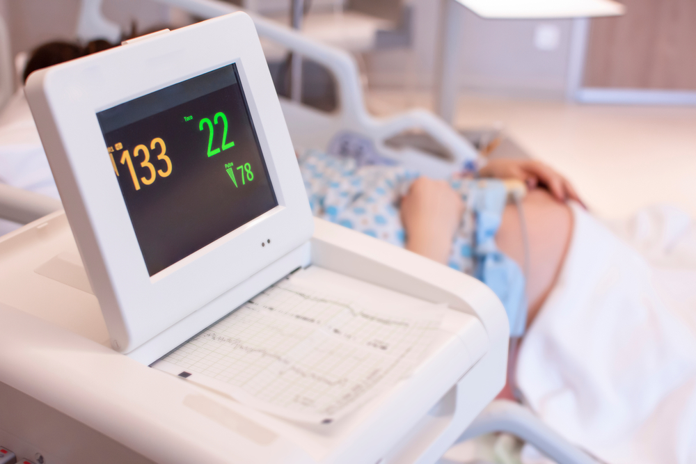
The primary equipment used is an Electronic Fetal Monitor. The mother is typically in a reclined or left-lateral position. Two transducers (sensors) are secured to her abdomen with elastic belts:
The test is usually administered by a Labor and Delivery (L&D) Nurse or a specialized technician in an outpatient setting. However, the ultimate responsibility for “reading” and signing off on the strip lies with the doctors, such as a Obstetrician (OB/GYN) or a Maternal-Fetal Medicine (MFM) specialist. The provider must ensure the equipment is calibrated correctly and that the mother is positioned to avoid “supine hypotension,” which can artificially depress the baby’s heart rate.
The NST monitors the relationship between fetal movement and the heart rate. The monitor produces a continuous paper strip or digital graph showing the heart rate in beats per minute (BPM).
The goal of the NST is to determine if the fetus is “Reactive.”
| Result | Definition | Clinical Meaning |
| Reactive | Two or more heart rate accelerations of at least 15 BPM above baseline, lasting at least 15 seconds, within a 20-minute window. | Indicates a well-oxygenated fetus with a functioning neurological system. |
| Non-Reactive | Lack of sufficient accelerations over a 40-minute period (accounting for fetal sleep cycles). | Possible sign of fetal distress, hypoxia (oxygen deprivation), or sedation. |
A Reactive NST generally allows the mother to go home with instructions to continue monitoring fetal movement.
A Non-Reactive NST is a red flag. It does not always mean the baby is in danger—the baby could simply be sleeping—but it requires immediate “reflexive” testing. The provider should:
Medical malpractice occurs when a healthcare provider deviates from the “standard of care,” resulting in injury. In the context of NSTs, malpractice usually falls into two categories: the failure to perform a test and the failure to interpret/react to one.
If a mother presents with “red flag” symptoms—such as high blood pressure or decreased fetal movement—and the doctor fails to order an NST, they may be liable for any subsequent injury. For example, if a placenta is failing (placental insufficiency) and the doctor ignores the signs, the baby may suffer from chronic oxygen deprivation.
This is the most common form of NST malpractice. A nurse or doctor may misread a “Non-Reactive” strip as “Reactive,” or they may fail to notice decelerations (drops in heart rate). Late decelerations during an NST are particularly ominous, as they often signal that the baby is struggling to maintain oxygen levels.
Even if the test is interpreted correctly, a delay in response can be fatal. If a BPP is indicated but the doctor waits several hours to perform it—or waits too long to order an emergency C-section—the baby may suffer Hypoxic Ischemic Encephalopathy (HIE).
Sometimes, malpractice earlier in the pregnancy (like failing to manage gestational diabetes) creates a high-risk environment that necessitates frequent NSTs. If those tests are then managed poorly, it compounds the original negligence.
When an NST is mismanaged, the resulting lack of oxygen (asphyxia) can lead to permanent brain damage. Conditions often linked to NST malpractice include:
The Non-Stress Test is one of the most vital “windows” into the womb. When doctors and nurses look through that window, they have a legal and ethical duty to act on what they see. If your family has been impacted by a birth injury where fetal monitoring was a factor, it is essential to have the monitoring strips reviewed by medical experts to determine if the standard of care was met.
While the Non-Stress Test (NST) is a great first-line screening tool, it has limitations. Because it only looks at the fetal heart rate, it can sometimes produce “false positives”—indicating a baby is in trouble when they are actually just in a deep sleep cycle.
To get a clearer picture, doctors often “reflex” to a Biophysical Profile (BPP). Think of the NST as a quick check-up, while the BPP is an in-depth physical exam via ultrasound.
The primary difference is the scope of data. The NST measures one variable (heart rate), whereas the BPP measures five distinct markers of fetal health.
| Feature | Non-Stress Test (NST) | Biophysical Profile (BPP) |
| Method | External monitors (belts) on the abdomen. | Real-time Ultrasound + NST results. |
| Variables | Fetal Heart Rate (FHR) only. | Heart rate, breathing, movement, muscle tone, and amniotic fluid. |
| Duration | 20 to 40 minutes. | Up to 30 minutes of continuous ultrasound. |
| Primary Goal | To see if the heart accelerates with movement. | To assess the overall “wellness” and oxygenation of the fetus. |
A BPP is scored on a scale of 0 to 10. Each of the following categories is worth 2 points if present and 0 points if absent:
The BPP is essential because of how the fetal brain reacts to a lack of oxygen (hypoxia). The parts of the brain that control these movements are sensitive to oxygen levels. As oxygen drops, the baby stops performing these activities in a specific order:
If a BPP shows a baby has stopped breathing or moving, it is a medical emergency.
In legal cases involving birth injuries, the BPP is often a “smoking gun.” Malpractice frequently occurs when:
When a Non-Stress Test (NST) comes back as “Non-Reactive,” it is a pivotal clinical moment. As a patient or a family member, your role shifts from passive observer to active advocate. Because the window for preventing a birth injury like HIE can be narrow, asking the right questions—and demanding clear answers—is essential.
Here is a checklist of high-priority questions to ask your medical team if a “Non-Stress” result is concerning:
If you asked these questions and the medical team dismissed your concerns—only for your child to be born with a brain injury or requiring a NICU stay—it is time to look at the Electronic Fetal Monitoring (EFM) strips. These strips are the “black box” of the delivery room; they provide an objective, timed record of exactly what the baby was experiencing and whether the doctors ignored the warning signs.
To understand why medical malpractice often hinges on a fetal heart rate strip, you have to look at variability. While accelerations (the “stars” of the NST) show the baby is moving, variability shows the baby’s brain and nervous system are actively communicating with the heart.
In the legal world, variability is often the “canary in the coal mine.” If a doctor ignores “flat” or “minimal” variability, they are often ignoring the earliest signs of fetal brain hypoxia.
Variability refers to the natural, beat-to-beat fluctuations in the fetal heart rate. A healthy baby’s heart rate is not a steady, flat line like a metronome; it should look “jagged” or “shaggy” on the monitor.
These tiny jumps and drops (measured in beats per minute) are caused by the constant “push and pull” between the baby’s sympathetic and parasympathetic nervous systems.
Medical providers categorize what they see on the strip into four distinct levels:
In a Non-Stress Test, a baby can be “Non-Reactive” (no accelerations) but still be okay if they have Moderate Variability. However, if a baby is Non-Reactive and has Minimal or Absent Variability, the situation is dire.
Variability is the best indicator of “fetal reserve”—the baby’s ability to withstand the stress of labor. When variability disappears, it means the baby’s brain is no longer able to regulate the heart rate, often because it is diverting all available oxygen to vital organs.
In birth injury litigation, experts look for a “Category II” or “Category III” fetal heart rate tracing. Malpractice often occurs in the following ways:
Unlike a dramatic heart rate drop (bradycardia), loss of variability can be “silent.” It requires a trained eye to notice that the heart rate has become too “smooth.” If a provider misses this subtle shift, the window to perform a life-saving C-section may close, leading to permanent brain damage.
While variability shows the baby’s “reserve,” decelerations are the actual alarms going off on the monitor. A deceleration is a drop in the fetal heart rate below the baseline.
In a medical malpractice context, the timing of the drop in relation to the mother’s contractions is what determines if the baby is safe or in immediate danger.
Obstetricians categorize these drops into three main buckets: Early, Variable, and Late.
In rare and tragic cases, the monitor shows a “Sinusoidal” pattern—a smooth, undulating sine wave that persists for 20 minutes or more. This is not technically a deceleration, but a total breakdown of variability. It often indicates severe fetal anemia or massive internal bleeding (fetal-maternal hemorrhage). Ignoring a sinusoidal pattern is almost always a gross deviation from the standard of care.
A single “late” or “variable” deceleration isn’t necessarily malpractice, but a pattern of them is a mandate for action. Malpractice occurs when:
You can read a Baltimore Medical Malpractice Lawyer Blog post on a verdict involving decelerations, Fetal Decelerations $29M, and also other birth injury verdicts.
| Feature | What it tells the Doctor |
| Accelerations | The baby is awake and reactive. |
| Variability | The baby’s brain and heart are communicating well. |
| Decelerations | The baby is under stress (Head, Cord, or Placenta). |
When these signals are misread, the “window of opportunity” to deliver a healthy baby closes.
In a birth injury medical malpractice case, a “Theory of Liability” is the roadmap a legal team uses to prove that a healthcare provider’s actions (or lack thereof) caused a child’s injury. It connects the clinical data we’ve discussed—the NST, BPP, variability, and decelerations—into a single narrative of negligence.
To win a case, an attorney must prove four elements: Duty, Breach, Causation, and Damages. Here is how those elements typically come together:
The core of most NST/fetal monitoring lawsuits is the Breach of the Standard of Care. This rarely happens because of one single “bad” heartbeat. Instead, it’s a failure to see a downward trend.
Causation is often the hardest part to prove. The hospital’s defense will often argue that the brain injury happened before the labor or a genetic condition caused it, not the monitoring.
Often, a birth injury isn’t the result of one mistake, but a series of small ones that “line up” like holes in slices of Swiss cheese:
When a legal expert reviews a case for malpractice, they are looking for the “Triple Threat” of evidence:
If all three are present on the strip and the doctor does not deliver the baby immediately, the “Theory of Liability” is very strong.
Because these strips are complex, a lawsuit requires Expert Witnesses—usually board-certified OB/GYNs and Labor & Delivery nurses. They recreate the timeline, beat by beat, to show the jury exactly where the medical team “fell asleep at the wheel.”
If you have any concerns or questions about non-stress test medical malpractice and birth injury, then visit the Kopec Law Firm free consultation page or video. Then contact us at 800-604-0704 to speak directly with Attorney Mark Kopec. He is a top-rated Baltimore medical malpractice lawyer. The Kopec Law Firm is in Baltimore and pursues cases throughout Maryland and Washington, D.C.